ECtopic Fallopian
Tube Case
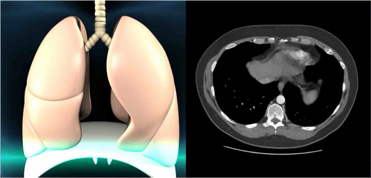
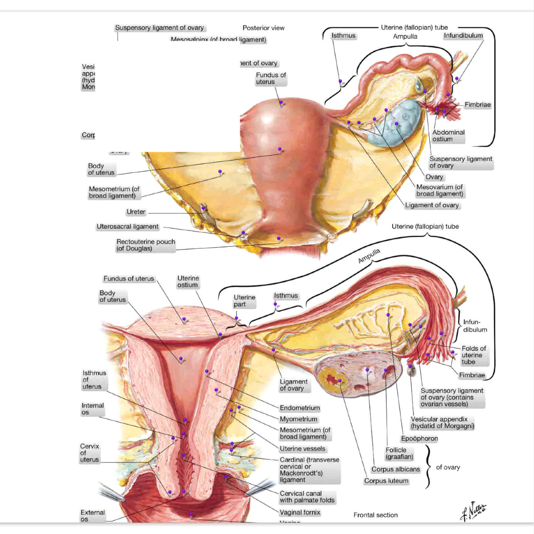
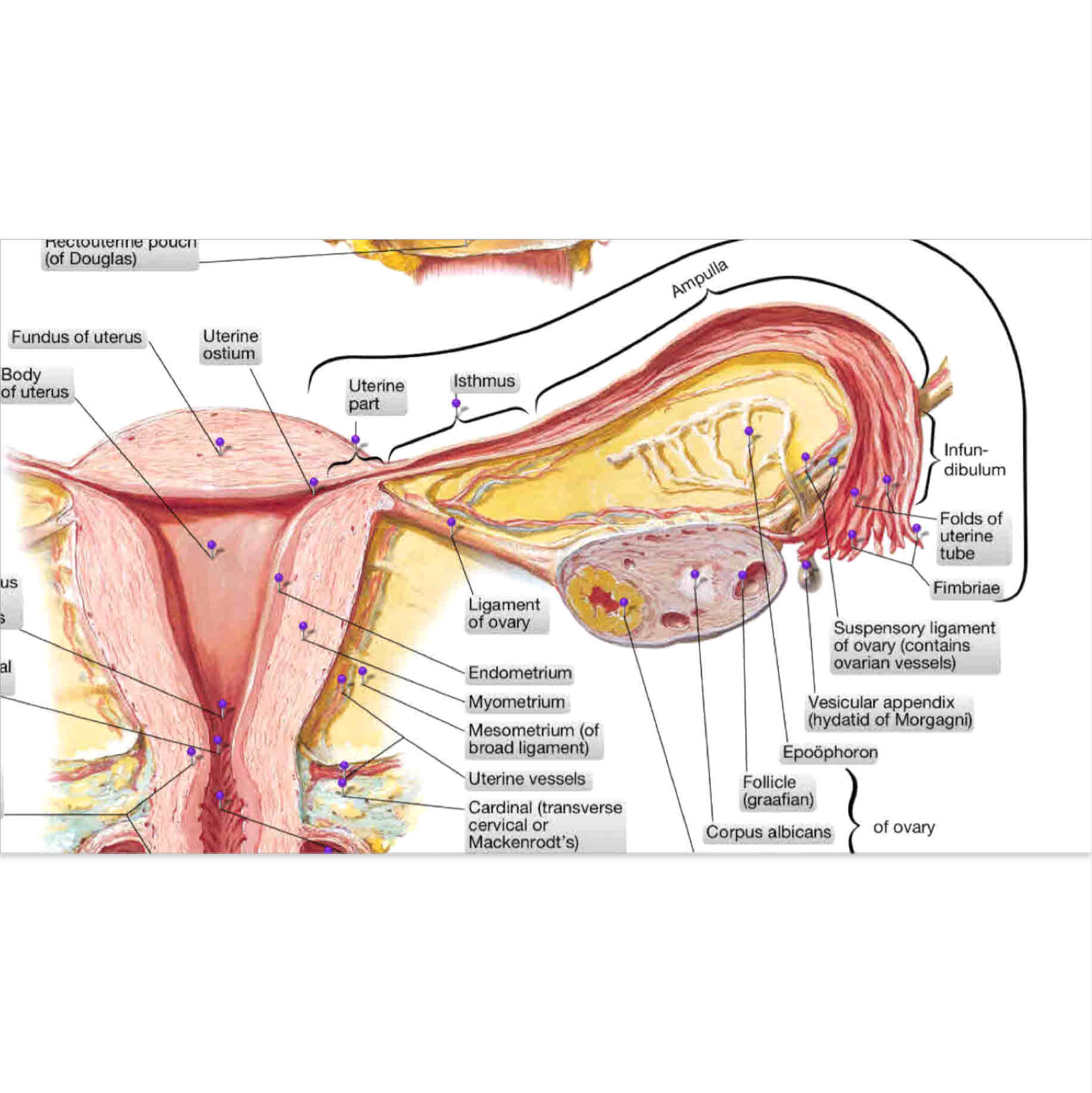
Plaintiff, an unmarried woman but with a steady boyfriend, presented to defendants fetal medicine M.D. for a staging sonogram at 9 weeks gestation. She had already undergone a right salpingectomy and right oopherectomy (image 1 shows the pre-treatment state with missing right fallopian tube and ovary-important to damages as plaintiff lost her left tube due to the malpractice and was left with only a left ovary such that future fertility requires IVF approach).
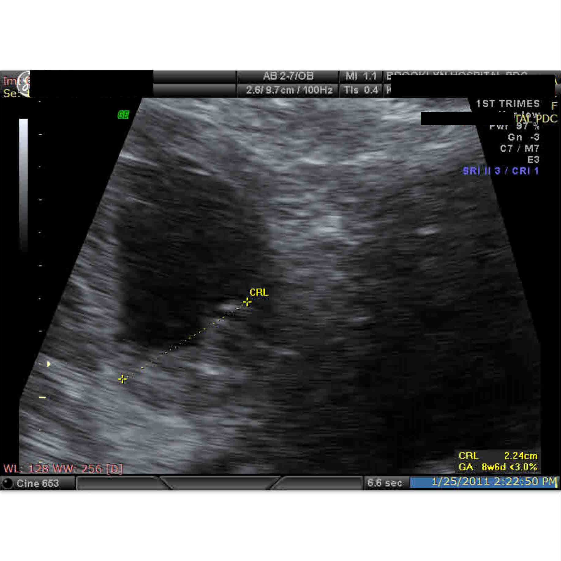
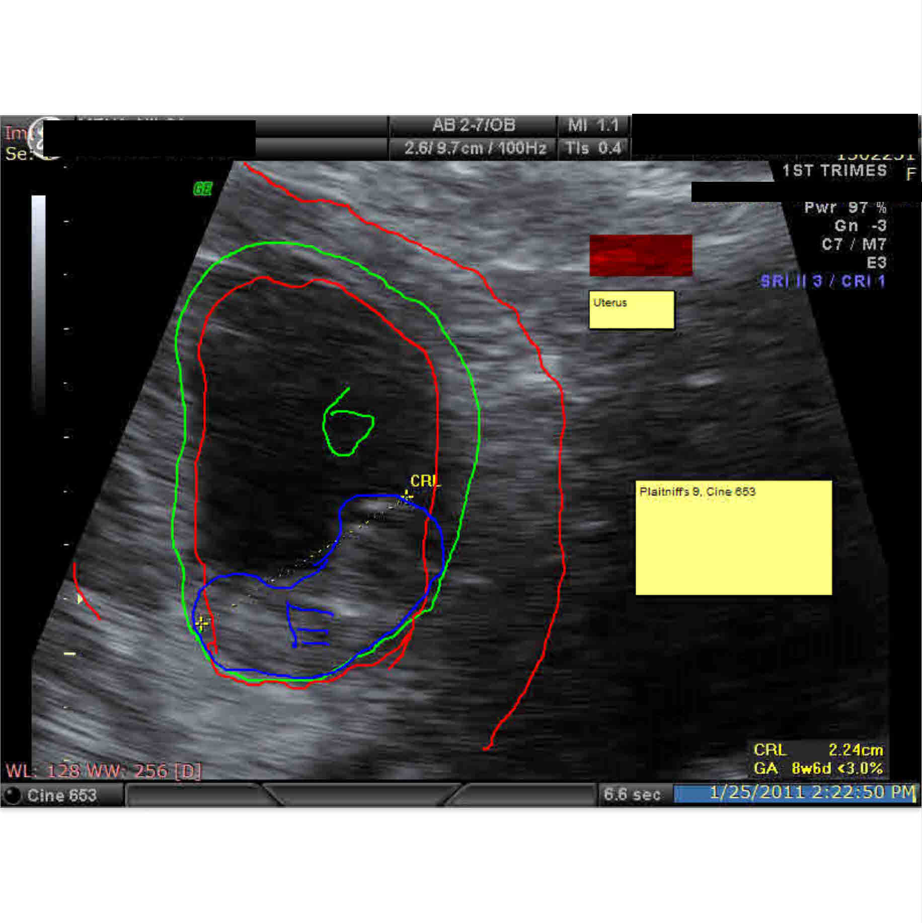
She reported bleeding and spotting with abdominal pain yet this history was not recognized as significant as possible indications of an ectopic pregnancy. 13 Sonogram images where made, electronically pre-marked at pretrial deposition, one of which was marked electronically live in front of the jury during trial by defendant himself (image 5 and 6 here, 6 showing the markings made by defendant with the red structure, by his erroneous view, showing the uterus which in truth demonstated an ectopic pregnancy in the left fallopian tube.
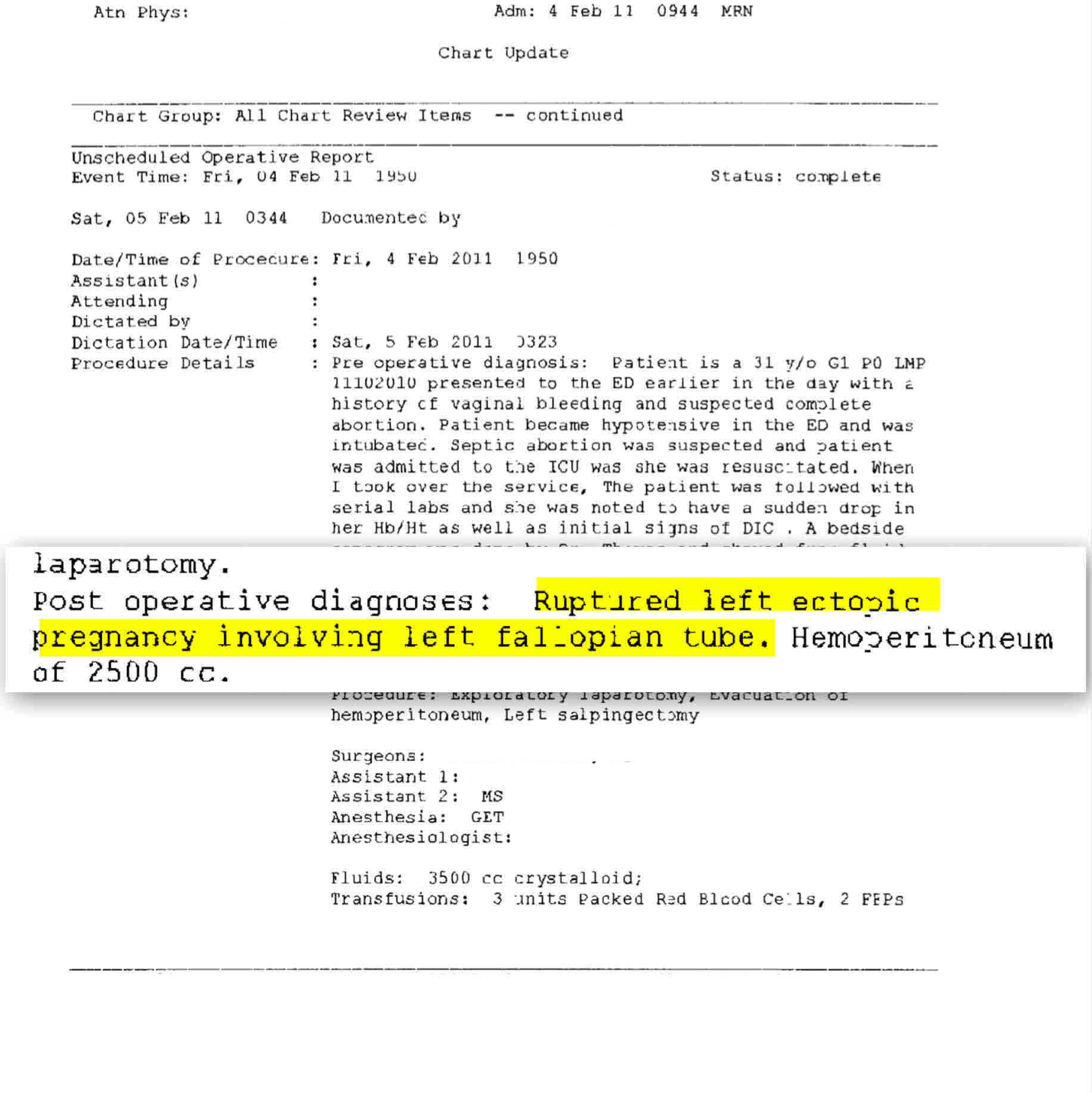
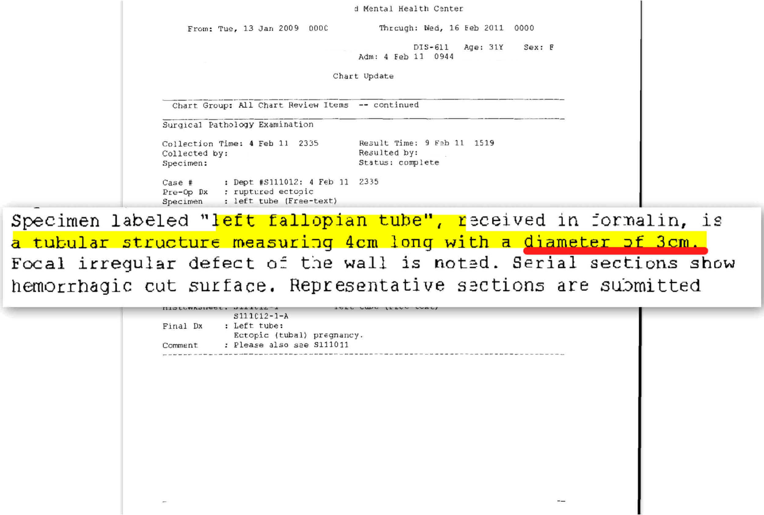
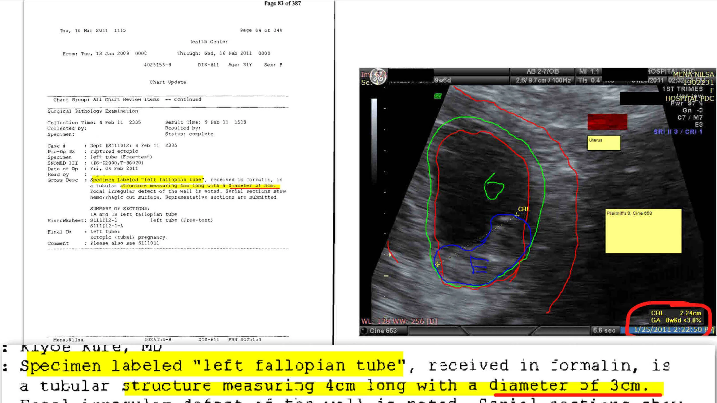
Defendant claimed that this was an interstitial pregnancy (at or about the junction between the uterus and fallopian tube) and not a tubal pregnancy. The crown-rump length noted on the subject image (2.24 cm, bottom left of images 5 and 6 here) clearly demonstrated that while defendant claimed that the uterus (in red by his own one-the-stand at trial marking) contained the pregnancy, in fact the size of the ruptured tube taken during the emergency surgery (3 cm in diameter) matched the size of the claimed uterus-which was in fact the fallopian tube by measurement.
Thus, with the imaging techniques of YourTrialSupport.com, yielded a plaintiff-favorable jury finding which went to make substantial award to plaintiff.
YourTrialSupport.com can assist you to a favorable result through formulating and sequencing persuasive visual evidence which keeps a jury fully engaged.